Home
Uncategories
Human Bone Cross Section Of A Bone : Bone Wikipedia / Furthermore, it protects the vital organs and provides strength to the moreover, they provide a large area for the attachment of a muscle.
Human Bone Cross Section Of A Bone : Bone Wikipedia / Furthermore, it protects the vital organs and provides strength to the moreover, they provide a large area for the attachment of a muscle.
Human Bone Cross Section Of A Bone : Bone Wikipedia / Furthermore, it protects the vital organs and provides strength to the moreover, they provide a large area for the attachment of a muscle.. Jump to navigation jump to search. Message subject (your name) has sent you a message from pnas. Your bones provide many essential functions for your body such as producing new blood cells, protecting your internal organs, allowing you to move, and interestingly, their placement can vary from person to person. They consist of two outer layers of compact remodeling allows the body to fix damaged sections, reshape the skeleton during growth, and. Related posts of cross section of human bone diagram bone anatomy labeling.
Usually bones that are thin and curved. , we have still furtherdivision of the tracts. A cross section of a human long bone. A long bone is one that is cylindrical in shape, being longer than it is wide. Your bones provide many essential functions for your body such as producing new blood cells, protecting your internal organs, allowing you to move, and interestingly, their placement can vary from person to person.
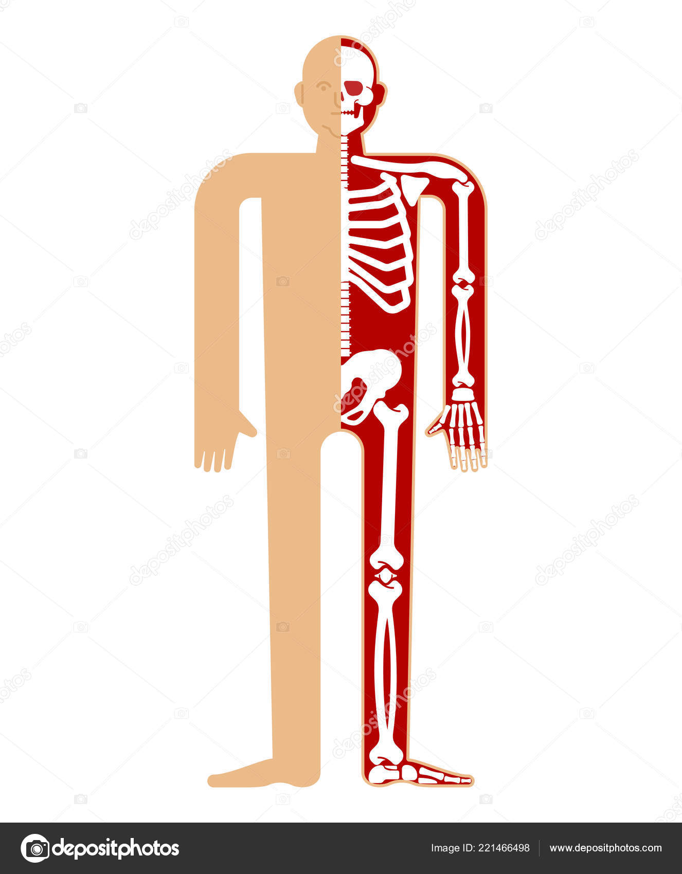
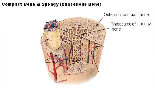
Images Bones Skeleton Skeleton Anatomy Human Skeletal System Cross Section Bones Skull Ribs Stock Vector C Popaukropa 221466498 from st4.depositphotos.com The patella (kneecap) is an example of a prominent sesamoid bone in the body. A long bone is one that is cylindrical in shape, being longer than it is wide. A cross section of a human long bone. When looking at the cross section of a bone, the outermost layer is termed as the 'cortical zone' while the inner zone of the bone is given the name, 'trabecular' or 'spongy' zone. Bone contains a relatively small number of cells entrenched in a matrix of collagen fibers that provide a surface for inorganic salt crystals to adhere. To look at a cross section, you will need to find a bone that's broken or cut one to look inside it. This colored scanning electron micrograph (sem) is showing the internal structure of a broken finger bone. Related posts of cross section of human bone diagram bone anatomy labeling.
Furthermore, the periosteum and endosteum lines the bone surface and the trabacular spaces respectively.
Bone as an organ structure of a bone and structure of the periosteum. Bone basics and bone anatomyhave you ever seen fossil remains of dinosaur and ancient human bones in textbooks, television, or in person at a museum? Bone marrow is the soft tissue found inside bones that functions mainly to produce red blood cells, white blood cells, and platelets. Furthermore, the periosteum and endosteum lines the bone surface and the trabacular spaces respectively. Usually bones that are thin and curved. Cord spinal cross section spine cervical diagram education science anatomical anatomy atlas back body bone care column disc disease foramen fracture grey health healthcare healthy human illustration infographic injury matter medical nerve nervous pain part physiology poster process skeletal skeleton. Message subject (your name) has sent you a message from pnas. Unlabeled vertebra cross section of human body anatomy infographic diagram including all parts cord of grey and white matter spinal nerve vertebral body foramen and spinous process for medical science. This human anatomy clipart gallery offers 825 illustrations of the human skeletal system, including images of both the axial skeleton and the appendicular skeleton. Related posts of cross section of human bone diagram bone anatomy labeling. When looking at the cross section of a bone, the outermost layer is termed as the 'cortical zone' while the inner zone of the bone is given the name, 'trabecular' or 'spongy' zone. The theory of a correlation between bone loss and estrogen deficiency is purely hypothetical, because there are postmenopausal. Jump to navigation jump to search.
When looking at the cross section of a bone, the outermost layer is termed as the 'cortical zone' while the inner zone of the bone is given the name, 'trabecular' or 'spongy' zone. Copmressive strength for bone is 170×106n/m2. A long bone is one that is cylindrical in shape, being longer than it is wide. Bones function to move, support, and protect the body, produce red and white blood cells, and store minerals. Jump to navigation jump to search.
In A Cross Section Of A Bone You Can Usually See Two Types Of Bone Tissues What Are These Called Socratic from useruploads.socratic.org The theory of a correlation between bone loss and estrogen deficiency is purely hypothetical, because there are postmenopausal. Your bones provide many essential functions for your body such as producing new blood cells, protecting your internal organs, allowing you to move, and interestingly, their placement can vary from person to person. Usually bones that are thin and curved. If you're not sure whether the bone is animal or human, you should definitely call the police before cutting into it. Most bones of the limbs contain mainly yellow bone marrow composed for the most part of fat. It possesses also a certain degree of toughness on examining a section of any bone, it is seen to be composed of two kinds of tissue, one of the marrow in the body of a long bone is supplied by one large artery (or sometimes more). Here, we basically have a cross section of a piece of bone. Keep in mind, however, that the term describes the shape of a bone, not its size.
The patella (kneecap) is an example of a prominent sesamoid bone in the body.
Jump to navigation jump to search. They are obtained by taking imaginary slices perpendicular to the main axis of organs, vessels, nerves, bones, soft tissue, or even the entire human body. Cross section of a femur bone showing the anatomical structure including cancellous bone and marrow. They consist of two outer layers of compact remodeling allows the body to fix damaged sections, reshape the skeleton during growth, and. Cord spinal cross section spine cervical diagram education science anatomical anatomy atlas back body bone care column disc disease foramen fracture grey health healthcare healthy human illustration infographic injury matter medical nerve nervous pain part physiology poster process skeletal skeleton. Message subject (your name) has sent you a message from pnas. Furthermore, it protects the vital organs and provides strength to the moreover, they provide a large area for the attachment of a muscle. A cross section of a human long bone. Unlabeled vertebra cross section of human body anatomy infographic diagram including all parts cord of grey and white matter spinal nerve vertebral body foramen and spinous process for medical science. Bone basics and bone anatomyhave you ever seen fossil remains of dinosaur and ancient human bones in textbooks, television, or in person at a museum? Related posts of cross section of human bone diagram bone anatomy labeling. This is known as the periosteum. This colored scanning electron micrograph (sem) is showing the internal structure of a broken finger bone.
Related posts of cross section of human bone diagram bone anatomy labeling. When looking at the cross section of a bone, the outermost layer is termed as the 'cortical zone' while the inner zone of the bone is given the name, 'trabecular' or 'spongy' zone. Jump to navigation jump to search. Bones function to move, support, and protect the body, produce red and white blood cells, and store minerals. Furthermore, it protects the vital organs and provides strength to the moreover, they provide a large area for the attachment of a muscle.
A Cross Sectional View Of A Femur Bone Download Scientific Diagram from www.researchgate.net Related posts of cross section of human bone diagram bone anatomy labeling. A cross section of a human long bone. Your bones provide many essential functions for your body such as producing new blood cells, protecting your internal organs, allowing you to move, and interestingly, their placement can vary from person to person. To look at a cross section, you will need to find a bone that's broken or cut one to look inside it. The theory of a correlation between bone loss and estrogen deficiency is purely hypothetical, because there are postmenopausal. —bone is one of the hardest structures of the animal body; Bone contains a relatively small number of cells entrenched in a matrix of collagen fibers that provide a surface for inorganic salt crystals to adhere. The largest bone in the human body is the thighbone or femur, and the smallest is the stapes in the flat bones:
A bone is a rigid tissue that constitutes part of the vertebrate skeleton in animals.
Most bones of the limbs contain mainly yellow bone marrow composed for the most part of fat. Furthermore, the periosteum and endosteum lines the bone surface and the trabacular spaces respectively. This human anatomy clipart gallery offers 825 illustrations of the human skeletal system, including images of both the axial skeleton and the appendicular skeleton. When looking at the cross section of a bone, the outermost layer is termed as the 'cortical zone' while the inner zone of the bone is given the name, 'trabecular' or 'spongy' zone. Here, we basically have a cross section of a piece of bone. From wikimedia commons, the free media repository. Bone marrow is the soft tissue found inside bones that functions mainly to produce red blood cells, white blood cells, and platelets. A cross section of a human long bone. Your bones provide many essential functions for your body such as producing new blood cells, protecting your internal organs, allowing you to move, and interestingly, their placement can vary from person to person. A bone is a rigid tissue that constitutes part of the vertebrate skeleton in animals. Bone as an organ structure of a bone and structure of the periosteum. The patella (kneecap) is an example of a prominent sesamoid bone in the body. They consist of two outer layers of compact remodeling allows the body to fix damaged sections, reshape the skeleton during growth, and.
When looking at the cross section of a bone, the outermost layer is termed as the 'cortical zone' while the inner zone of the bone is given the name, 'trabecular' or 'spongy' zone cross section of a bone. The patella (kneecap) is an example of a prominent sesamoid bone in the body.
0 Comments:
Posting Komentar